18F-DOPA PET/CT imaging in endocrine disorders - The Australian experience (#73)
Introduction: 18F-3,4-dihydroxyphenylalanine Positron Emission Tomography with Computed Tomography (18F-DOPA PET/CT) is a novel technique in the assessment of endocrine tumours and congenital hyperinsulinism of infancy (CHI). 18F-DOPA PET/CT has demonstrated promise in superior diagnostic performance in limited clinical studies internationally. Experience from the only centre in Australia offering 18F-DOPA is described.
Methods: Retrospective evaluation of data from all patients who underwent 18F-DOPA PET/CT was performed. Patients were generally considered eligible for an 18F -DOPA scan if there was strong suspicion or proven biochemical evidence of neuroendocrine tumour, but negative or equivocal conventional imaging tests. Adult patients received approximately 200mBq of 18F-DOPA while children received 6mBq/kg. A 30-60 minute uptake time was observed for all indications other than CHI. For the latter, dynamic imaging was performed over 60 minutes. Low-dose CT scan for anatomical correlation was also performed. Images were jointly reported by two experienced nuclear medicine specialists, with knowledge of previous imaging and biochemical findings.
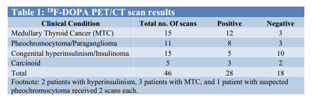
Results: 18F-DOPA PET/CT scan indications included diagnosis, characterisation and restaging of a number of endocrine tumours and CHI. 46 scans from 40 patients (age range 0-79 years), were included. Scan indications and results are summarised in the table below. Scans were positive in 61% (28/46) of studies, and these findings will be presented in detail.
Conclusion: Our initial results with 18F-DOPA imaging are promising, with 61% of patients showing abnormal 18F-DOPA uptake. Many of these patients had comprehensive conventional imaging work-up prior to 18F-DOPA scan and therefore, it is likely that 18F-DOPA imaging has potential to significantly impact the management of patients with endocrine tumours and CHI.

 ESA-SRB 2013*